Keywords: Protease inhabitor treatment, HIV viral, tonsil lymphatic tissue, T-lymphocyte, influencza, clearing up herpes outbreaks, immune system stimulation, vitamine C, minerals, CD4, AZT8, DDI, painful lesions, ZDV, DDI, DD3, 3TC, AZT, lyphatic fluids, tissues, Short-Wave Diathermy, PHA(phytohaemagglutinin), (PEMF)Low-frequency pulse electromagnetic field, THERMASTIM, Viral Treatment System, protease inhibitors, T-cell,
Part 1 of 4
March 14, 1998
To : P.T.Pappas
Enclosed is a copy of the AIDS IDC that I prepared for Gregg. I am quite sure I had given you a copy of this in Minnesota. Nevertheless I here send it to you again.
Sincerely yours
Paul A. La Violette
Investigational Device Exemption
PAP Electrodynamics, USA
8793 Topanga Canyon Blvd., Suite 111
Canoga Park, CA 91304
PAP Electrodynamics Agency
1176 Hedgewood Lane
Niskayuna, NY 12309
Index |
|
| A. | Purpose |
| B. | Justification |
| C. | Prior Investigations |
| 1. Results of PAP IMI Treatment of AIDS Patients | |
| 2. Electroinsertion of CD4 Molekules as a Possibile Explanation of the PAP IMI Results | |
| 3. Electric Current Inactivation of the AIDS Virus | |
| 4. Other Research Indicating Vital Lysis by High Frequency Electric Fields | |
| 5. Treatment 0f AIDS Patient with Other Pulsed EM Field Devices | |
| D. | Description and Operation of the Device |
| 1. Device Description | |
| 2. Principle of Operation | |
| 3. Advantage of the Pulse Brivity of the PAP IMI Compared with Other Diathermy Device | |
| 4. Labeling for the PAP IMI | |
| E. | Risk Analysis |
| 1. Electromagnetic Radiation Comparison to Surgical Diathermies | |
| 2. Reports of the Adverse Consequences | |
| 3. Animal Studies | |
| 4. Reports of Favorable Treatment Results | |
| 5. Precautions | |
| 6. Contradictions for Special Medical Conditions (applicable to all diathermies) | |
| 7. Safety Features | |
| 8. Low Risk to High-Voltage Exposure | |
| F. | Protocol |
| 1. Study Overview | |
| 2. Personnel and Institutions | |
| 3. Criteria for Patient selection and for Exlusion of Patients and an Estimate of the Number of Patients to be Studied | |
| 4. Patient Confidentiality | |
| 5. Informed Consent | |
| 6. Invesigationnal Review Board | |
| 7. Methodology | |
| 8. References | |
| 9. Appendices | |
Investigational Device Exemption
A. PURPOSE
The purpose of this study is to investigate the safety and efficacy of treating HIV positive patients with the PAP Ion Magnetic Inductor (IMI) diathermy device.
Great steps forward have been made in the pharmacological
treatment of AIDS through the use of combination therapy treatments incorporating protease
inhibitors. Patients who have followed the protease inhibitor treatment protocols for a
sufficiently long period of time have been found to have HIV viral load levels in their
blood that fall below the level of detection. Nevertheless, while such treatments may
suppress viral load in the blood, some form of the virus may still remain in a potentially
infective state. For example, one study found HIV DNA present in tonsil lymphatic tissue
of patients taking protease inhibitor combination therapy for 24 weeks even though their
blood viral load levels had declined dramatically (Science, val. 275, Jan. 31,
1997, p. 616). So it is believed that protease inhibitors may at best keep the HIV
infection in check only as long as their consumption is continued. But because the cost of
these antiviral medications is very high, the expense of their extended use can be
overwhelming for most uninsured individuals.
We wish to investigate the clinical effectiveness of an alternative
method of AIDS treatment that has been found to produce antiviral effects more dramatic
than those obtained with protease inhibitors, and far less costly. This therapy involves
use of the PAP IMI to apply very high-intensity, AC magnetic field pulses to the patient's
body. Specific target areas for treatment include the lymph nodes, which are known to
preferentially harbour the HIV virus especially in the early stages of the disease, the
spleen, which has a key role in filtering the blood, and the thymus, which is a key link
in the immune response to fighting the disease through its production of CD4 cells.
Past experience with the PAP IMI has shown that its effects on AIDS
patients are to dramatically reduce HIV viral load as well as dramatically increase
T-lymphocyte count toward normal levels. Thus the effects of the PAP IMI appear to be both
viral destroying and immunostimulatory, although the two effects are undoubtedly
intertwined. Compared with protease inhibitor therapy, the PAP IMI treatment is capable of
producing far more dramatic results. For example, unaided by antiviral medication, it has
induced CD4 cell count increases of as much as 240 ml-1 within just a one month period and
as much as 850 min in just a 7 month period. By comparison, the protease inhibitor
nelfinavir0 in combination with AZT and 3TC was found to produce a rise in CD4 cell count
of only 155 to 160 m1'1 after 6 months of treatment.
PAP IMI treatments are initially administered hi-weekly, and are
eventually decreased to semi-monthly or monthly maintenance treatments as the patient's
condition improved. Moreover there are indications that the treatment can eventually be
discontinued. For example, one AIDS patient, who received combination treatments with the
PAP IMI and with a micro current device for approximately 1-1/2 years, maintained a blood
viral load level below detection a full 10 months after the therapy was discontinued and
without the help of antiviral medication. If future viral load tests of this patient show
similar results" this may be an indication that the PAP IMI combination therapy is
capable of completely eradicate the virus.
Let us first take a moment to review past research that 1) is
relevant to the safety and effectiveness of the PAPIMI device for use in treating HIV
infections and AIDS and 2) that justifies the need for carrying out the proposed research
study.
1. Results
of PAP IMI Treatment of AIDS Patients.
a) Initial discovery of PAP IMI efficacy for AIDS treatment and early results:
Dr. Jacob Swilling, M.D., of Mexico was the first to recognise in
1992 that PAP IMI treatments might be beneficial to the treatment of AIDS. This indication
was further explored in the following year at the International Pain Research Institute of
Beverley Hills, CA where a small number of AIDS patients received treatment with the PAP
IMI. Appendix A presents testimonials given by three of these AIDS patients. CD4 data,
available for one of these patients shows that his CD4 count increased an astounding 19
fold over the course of his 9 month treatment period; see Figure l.
Also, about this same time other evidence came to light which also
indicated that the PAP IMI may have immunostimulatory or antiviral effects. For example,
Dr. Sam Chachoua of ------- in ----------- found that PAP IMI exposures administered after
vaccination to 21 patients stimulated antibody production. Also there were anecdotal
reports that PAP IMI treatments was useful in preventing or stopping influenza, and in
temporarily clearing up herpes outbreaks.
 |
| Figure 1. Response of
T-cell count to PAP IMI treatment. Patient: HIV positive, male, 37 years old. Treatment
with PAP IMI: 20 minutes per week reducing to 20 min. every other week. Location: National Pain Institute (Santa Monica, CA) |
b) AIDS study conducted by Dr. Nick Tsilimigakis of the Scientific Institute for Bioenergy in Athens (Glyfada), Greece (mid 1994 to present):
Dr. Tsilimigakis has reported data on seven patients
receiving PAP IMI treatments over periods of time ranging from one month to a year. His
treatment protocol and the case history summaries of these patients are presented in
Appendix B. 1 Treatments
usually lasted for 20 minutes and were given two times per week. The probe was positioned
over the thymus, for immune system stimulation, as well as on infected regions or
problematic areas. This magnetic induction therapy was carried out in conjunction with a
pulsed DC micro current therapy in which 2 to 3 milliamperes of pulsed DC current at 45
volts were conducted through the patient, from one hand to the other. Each of these
treatments lasted about half an hour and were carried out two times a week. Usually large
doses of vitamin C and minerals were prescribed for the patients as well.
The Tsimilimigakis method obtained astounding results with a 100%
response. In all 7 cases, patients experienced immediate improvement in their physical
condition (i.e., more energy, better feeling, weight gain, alleviation of diarrhea
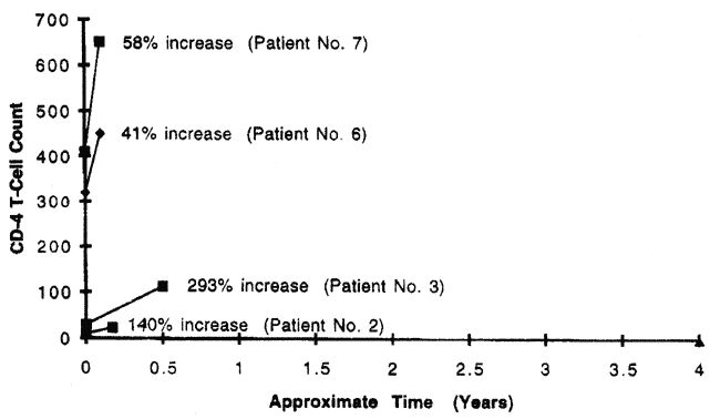
symptoms) followed by a precipitous increase in their CD4 cell counts, ranging from 41% in
one month (Case 6) to 293% in six months (Case 3); see Figures 2 and 3. In two cases the
initial CD4 cell counts of the patients were as low as 10 ml-1 and 30 ml-1. Only one of
these patients (Case 5) had been taking drug therapy (AZT & DDI) prior to treatment.
 |
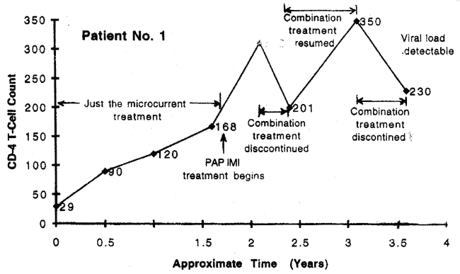
| Figure 2. Patient No.1: CD4 cell counts before and after treatment with the PAP IMI using the Tsilimigakis protocol. |
 |
| Figure 3. Patients Nos. 2, 3, 6, and 7: CD4 cell counts before and after treatment with the PAP IMI using the Tsilimigakis protocol. |
Although the PAP IMI was used in
combination with the micro current therapy technique, Dr. Tsilimigakis found that the PAP
IMI significantly boosted the therapeutic results, compared to just using the micro
current technique alone. Evidence of this is also seen in the CD4 count data in Case 1,
where a doubling of the rate of increase of the CD4 cell count is evident following
addition of the PAP IMI treatments to the micro current treatment regimen. That is, prior
to the beginning of combination treatment with the PAP IMI an increase of 6.7% per month
is evident in CD4 count, whereas after the introduction of combination treatment an
increase of 15.6% per month is evident.
In Case 1, the evidence suggests that the disease was completely
eradicated after 15 months of receiving PAP IMI along with micro current treatments, to
the extent that viral load measurements were unable to detect the HIV virus. Earlier in
the patient's treatment, there was an indication that the disease had not been completely
eradicated, but only counteracted. That is, after 4 months of PAP IMI treatment when the
patient discovered that her CD4 count had reached 312, she was so encouraged by the
results that she discontinued therapy to take a trip to France. However, after 4 months
without treatment her CD4 cell count had fallen 36%. Subsequently she resumed the
combination treatment and her count rose to a new high value. Again she discontinued
treatment and 10 months later found that her viral load was undetectable.
c) AIDS study conducted at the First Hospital of Social Security
(IKA) in Athens (Mellisia), Greece (February 1995 to present):
This study is being conducted by Drs. D. Stergiou, A. Papadopoulos,
J. Arkadianos, and A. Scoullos under the approval of the Greek National Drug Organization.
Dr. Stergiou reports data on 12 consecutive AIDS cases in which patients received PAP IMI
treatments over periods of time ranging from one to ten months (Appendix C). 2 Treatments
lasted 25 minutes and were given two times per week. The probe was positioned over the
spleen, the thymus, the axillary and neck lymph nodes, and in some cases over painful
lesions. Most of the patients were also taking some kind of retroviral medication (e.g.,
ZDV, DDI, DDC, 3TC, AZT). Other than this, the patients received no other
immunostimulatory therapy.
Very encouraging results were found. Four patients experienced a
dramatic increase in their CD4 cell counts, with increases ranging from 110% to 610% in
periods ranging from one to four months; see Figure 4. The CD4 counts of two other
patients remained stable during the three months that they underwent PAP IMI treatment
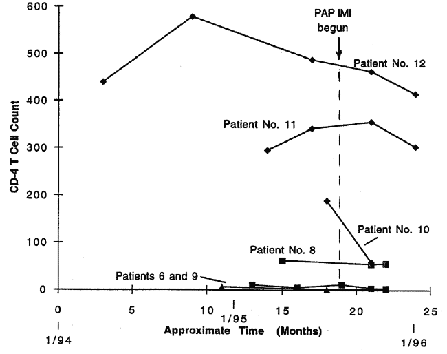
(Figure 5). Also the CD4 counts of three other patients decreased in the two to three
months that they underwent PAP IMI treatment (Figure 5). One of these latter three
(Patient 12) appears to have had CD4 count decreases that were already in progress prior
to PAP IMI treatment and which the treatment failed to counteract. So these decreases
should not be considered to be a result of the treatment. Patient No. 10, who experienced
a CD4 decrease of 68%, suffered from Kaposi's sarcoma, yet his lesions remained stable
during the treatment period. Patient No. 12, who experienced a CD4 decrease of 10%, was
diagnosed to be in excellent condition. In the third case (Patient No. 11), the decrease
was moderate, 15%, and despite the decrease, it was reported that the patient remained in
very good clinical condition.
 |
| Figure 5. Patient Nos. 6, 8, 9, 10, 1l., and 12: CD4 Cell counts before and after treatment with the PAP IMI in the IKA protocol. |
No CD4 count data are yet available on one of the
12 patients (Patient No. 7). Also out of this set of 12, there was one patient (No. 4)
whose condition worsened after receiving just five PAP IMI treatments. Before he could
receive the full benefits of the therapy, he had to discontinue treatment, and a month
later he died. No CD4 data is available in his case either. Unlike the other patients,
this patient was not taking retroviral medication at the time and his Kaposi's sarcoma was
rather advanced, with lesions being present in his lungs as well as on his skin.
Like the Tsilimigakis study, this study found that unless treatment
was continued for a sufficiently long period of time, gains in CD4 count could relapse if
the treatment was discontinued. This was seen in Patient No. 2. After attaining an
increase of her CD4 counts to 235 and continuing the PAP IMI treatments for a period of
only 3 months, the patient discontinued treatment, whereupon when measured some months her
CD4 count was found to have fallen five fold to 50.
CD4 count does not give the entire picture of the patient's response
to PAP IMI treatment. Viral load tests conducted on one patient showed that the patient's
viral load of HIV had decreased from 32,000 ml-1 to 22,000 ml-1 in the span of just one
month in which the patient had received just four PAP IMI treatments (once per week 20
minutes each). 3
Such a dramatic decrease in viral load and evidence mentioned in (b) above that at least
one patient had undetectable viral levels after just 15 months of treatment indicates that
the PAP IMI can be at least as effective as treatments using protease inhibitors.
The protocol used in the First Hospital of Social Security study and
case history summaries of the 12 patients are presented in Appendix C. Also the Appendix
includes an excerpt of Dr. Papadopoulos' conversation from the November 17, 1995
Hillman/PAP Electrodynamics teleconference.
2. Electro
insertion of CD4 Molecules as a Possible Explanation of the PAP IMI Results.
The PAP IMI generates in situ electric field intensities of the
order of 1.5 kV/cm during the first microsecond of every pulse discharge. This is
sufficiently high to induce several membranal phenomena. These include Electro
insertion - the insertion and permanent incorporation of molecules into the cell
membrane wall, electropermeability - the momentary increase in cell membrane
permeability during the duration of the pulse followed by a return to normal levels of
permeability, electrofusion - the fusion of juxtaposed cells during momentary
alteration of their membrane permeability. The electric field intensity threshold where
these effects begin to occur ranges from 0.3 to 0.6 kV/cm, which is exceeded by the PAP
IMI pulse at its initial peak intensity. 4
The PAP IMI peak field strength also lies at the lower end of the field strength range for
inducing membranal electroporation (1 to 10 kV/cm), electroporation involving the
temporary increase in membrane permeability resulting from the opening of membranal pores.
5
Experiments conducted on the electroinsertion phenomenon could
explain the elevated T cell counts observed in AIDS patients receiving PAP IMI treatments.
For example, Mouneimne et al. 6
have applied to human red blood cells in vitro a sequence of 4 electric field pulses
having a field intensity of 1.3 kV/cm and duration of a few milliseconds, with the pulses
spaced by a 15 minute rest period. Using this technique, they were able to insert up to
5000 CD4 molecules into each red blood cell membrane, while at the same time preserving
the immunological integrity of the proteins. These CD4 bearing erythrocytes (RBC-CD4) were
found to be able to internalize HIV-1 in vitro. Moreover the RBC-CD4 in vitro were found
to compete with T4 cells for HIV attachment. The preincubation of HIV-1 with RBC-CD4
reduced the infection of target cells by more than 90% as detected by viral reverse
transcriptase and the amount of P24 antigen produced by the target cells. The
electroinserted red blood cells exhibited a normal life span. The researchers theorized
that CD4 protein electroinserted into red blood cell membranes might provide a useful
therapeutic agent against AIDS. We believe that the PAP IMI pulses administered to AIDS
patients are electroinserting CD4 molecules present in the blood plasma into the patients'
red blood cells and thereby diverting the HIV-1 from its attack on CD4 cells. This would
result in the observed rise in CD4 cell count and observed decline in viral load.
3. Electric Current Inactivation of the AIDS Virus.
In March 1991, William D. Lyman and his colleagues
at the Albert Einstein College of Medicine (The Bronx, NY) reported research in which they
passed microcurrents through HIV contaminated blood. 8-9
They found that treatment durations as short as 6 minutes substantially incapacitated the
AIDS virus, halting its ability to reproduce.
In this study, 10 microliters of HIV-1 infected blood containing 105
infectious particles per ml were exposed to an electric current passing between two
platinum electrodes placed in direct contact with blood in vitro. Currents ranged from 25
to 100 microamperes (ia) and exposure times ranged up to 12 minutes. The researchers found
that exposing the virus to direct electric current suppressed its capacity to induce the
formation of syncytia, an indicator that quantifies the production of infectious
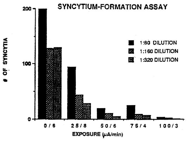
particles. Passing a total charge of 200 microamp minutes (25 ia for 8 minutes) through
the blood reduced the number of syncytia from 50 to 65% while a charge of 300 microamp
minutes (50 ia for 6 minutes) through the blood reduced the number of syncytia from 90%;
see Figure 6.g Also reverse transcriptase assay, an index of viral protein production, was
found to be negatively impacted. lZeverse transcriptase activity was almost totally
ablated (reduced by 94%) with an exposure to 100 ia for 6 minutes; see Figure 7.8 The
precipitous drop in both of these indicators with administration of increasing electric
charge (current X time) showed that viral infectivity was substantially impaired by this
electric current treatment. A reprint of this report is presented in Appendix E.
Also Steven Kaali has reported that in addition to inactivating the
AIDS virus, this microcurrent treatment also left their blood samples free of hepatitis.
This indicates that electric fields may be useful in inactivating other viruses besides
the AIDS virus. The blood cells themselves were unharmed by the treatment. 10
Subsequently, in August of 1992, Kaali obtained a U.S. patent
(#5,139, 684) on a device for treating blood with therapeutic microcurrents. 11
Applications for this include processing blood stored in blood banks to inactivate
potential viral contamination. Alternatively, a kind of dialysis machine was proposed that
would remove blood from the patient, inactivate potential viruses, and reintroduce it back
into the patient.
By comparison, the PAP IMI has the advantage of being able to noninvasively
induce microcurrents in vivo by means of magnetic induction. The high frequency
magnetic pulses emitted from the coil of its probe are able to induce therapeutic electric
fields in the patient's body to an effective depth of 10 to 15 centimeters, exposing not
only the patient’s blood, but also lymphatic fluids and tissues as well.
 |
| Figure 6. Syncytium - formation assay as a function of microcurrent exposure (Lyman, et al.). |
 |
| Figure 7. Reverse transcriptase activity as a function of microcurrent exposure (Lyman, et al.). |
4. Other Research
Indicating Viral Lisis by High Frequency Electric Fields.
a) Early diathermy findings:
In his book Short-Wave Diathermy, Tibor
Cholnoky cites work by Carpenter and Page which indicates that the increased heat
generated in the body by high frequency electromagnetic waves is unfavorable to viral
development. 12 They note
that the heat increases the rate of chemical processes associated with the body's immune
system response.
Cholnoky also cites research indicating that high frequency
electromagnetic waves can kill bacteria at ambient temperatures below those that would
normally be lethal for such bacteria (e.g., temperatures administered via a water bath).
As one explanation, they cite the focal heat theory which suggests that the "focal
heat" absorbed by a single bacterium may be higher than the average heat absorbed by
the body's tissues, in which case the bacterium itself would be exposed to lethal
temperatures.
b) Immune system stimulation by electromagnetic fields:
Several studies have found that electromagnetic fields to weak to
produce thermal effects, are nonetheless able to affect the growth and DNA synthesis of
many cell types, including lymphocytes. 13-19
Research by J. Walleczek on human lymphocytes has shown that EM fields can produce changes
in calcium transport and cause mediation of the mitogenic response (i.e., stimulation of
the division of cellular nuclei). 20 Also A. Cossarizza, et al. found that when
lymphocyte cultures were stimulated with PHA (phytohaemagglutinin), an extremely
low-frequency pulsed electromagnetic field (PEMF) caused a significant increase of cell
proliferation in lymphocytes from both young or old subjects, the effect being more
pronounced in cells from older people. 21
After PEMF exposure, cells from old people reached levels of 3H-TdR incorporation not
significantly different from those of unexposed cultures taken from young people. The
pulsed field consisted of a 2.5 mT magnetic field that every 20 ms was turned on for a
period of 2 ms. The field by itself, without the PHA stimulant, was not found to be
mitogenic. Also they found that this same pulsed magnetic field increased interleukin-2
utilization and interleukin-2 receptor expression on the plasma membrane in
mitogen-stimulated human lymphocytes taken from elderly subjects. 22 They found that IL-2 receptor expression
increased 5 to 23 percent in 10 out of 10 aged subjects. The percentage of activated T
lymphocytes also had increased markedly in 83 percent of these subjects.
These immune system research results lend further support our
finding that the high-frequency electromagnetic pulses produced by the PAP IMI stimulate
an increase in CD4 cell count in AIDS patients. Hence further investigation of PAP IMI
therapy on AIDS patients is warranted.
5. Treatment of AIDS Patients with Other
Pulsed EM Field Devices.
In late April of 1994, Physiodynamics Corporation instituted a
company funded research project on the treatment of AIDS, a study conducted under the
provisions of FDA Regulations permitting limited gathering of experimental data. For their
research the used the THERMASTIM0, an electromagnetic, neuromuscular stimulator authorized
for commercial distribution by the FDA.
The THERMASTIM produces a weak pulsed electric field administered
via electrodes attached to various locations on the body. The current was pulsed at
frequencies of 100, 500, and 10,000 pulses per second, at voltages ranging from 2.4 to 27
volts, at currents ranging from 7 to 96 milliamps, and at wattages ranging from 0.06 to
2.9 watts. An eight pad configuration was used to simultaneously stimulate the blood,
lymphoid systems and bone marrow, and a four pad configuration was used to treat the
spleen and thymus. Treatment times ranged from 10 to 45 minutes. Treatment frequency
ranged from every other day to once every fourth day. The protocol recommended that the
total body be treated for half the time and the spleen and thymus for the other half of
the time. Physiodynamics named this treatment protocol the VTS®, VTS standing for Viral
Treatment System. Supporting information on this treatment is presented in Appendix D.
Physiodynamics initially became interested in investigating the use
of the THERMASTIM® in treating AIDS patients following the announcement by Lyman et al.
of their findings that the AIDS virus could be inactivated by exposure to electric
currents.
According to one person involved in the VTS study, one AIDS patient
obtained several positive benefits during the first 7 months of treatment, such as
increased energy level, increased appetite, weight gain, and greater resistance to
infection (Appendix D-1). 23
However, there was no increase in his CD4 cell count, which was initially 3, later rose to
9, but finally fell to 1 by the seventh month of treatment: 3.0 (5/5/94), 3.0 (7/5/94),
5.0 (10/5/94), 3 (10/28/94), 9 (11/28/94), 1 (12/19/94); see Appendix D-2). 24 Also a study of six AIDS patients treated
over a 2 to 14 month period concluded that the treatment had no consistent effects on CD4,
CD8, or viral burden (Appendix D-3). 25
By comparison, the studies cited in Section 3 above indicate that
the PAP IMI has a far more beneficial effect on the immune system of AIDS patients.